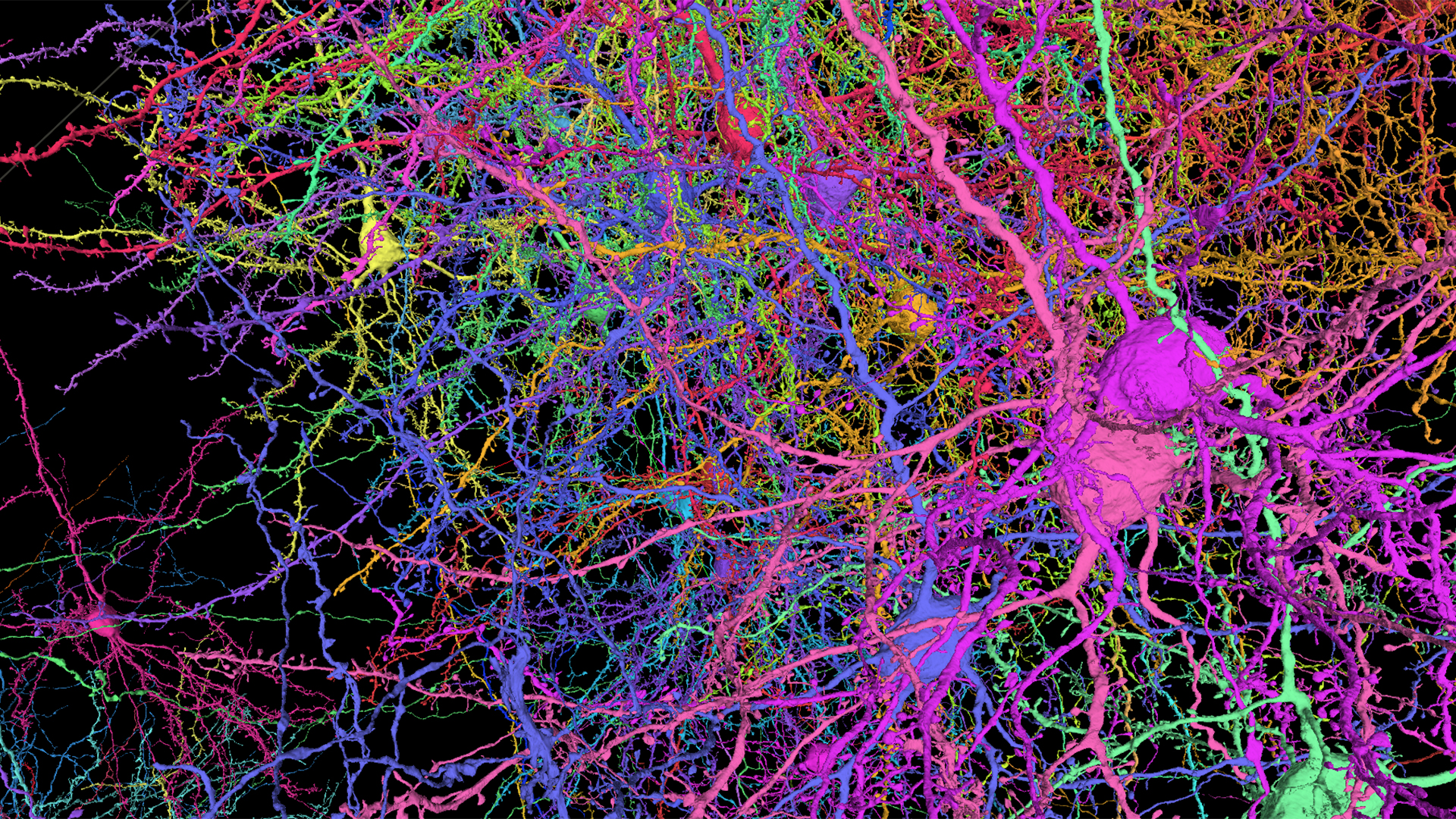
A 3-D reconstruction of cells from the MICrONS data set shows the complexity of the shapes and branched axons and dendrites in a mouse brain. Each cell is marked with a different color. (MICrONS / Allen Institutes)
Neuroscientists from the Allen Institute in Seattle and other research institutes completed a five-year, multi-million dollar project with the release of a high-resolution 3D map showing the connections between 200,000 cells in a lump of mouse brain about the size of a grain of sand.
The data collection, which is now publicly accessible online, was developed as part of the Machine Intelligence From Cortical Networks program, or MICrONS for short. MICrONS was funded in 2016 with federal grants of $ 100 million to the Allen Institute and its partners from the Intelligence Advanced Research Projects Activity, the US Secret Service’s equivalent to the Pentagon’s DARPA think tank.
MICrONS is set to pave the way for reverse engineering of the structure of the brain to help computer scientists create more human-like machine learning systems, but the database should also benefit biomedical researchers.
“We basically treat the brain circuit like a computer and asked ourselves three questions: What does it do? How is it wired? What is the program? “R. Clay Reid, senior researcher at Allen Institute and a senior scientist at MICrONS, said in a press release today.” Experiments were conducted to literally see neurons doing their math, to see them doing math . “
The newly released dataset includes 120,000 neurons as well as approximately 80,000 other types of brain cells, all contained within one cubic millimeter of the mouse brain’s visual neocortex. In addition to mapping the cells in physical space, the data set traces the functional connections of more than 523 million synapses.
Researchers from the Allen Institute were involved in the project by colleagues from Princeton University, Baylor College of Medicine, and other institutions.
Baylor’s team recorded the patterns of neural activity in a mouse while it was viewing images or films of natural scenes. After these experiments, the Allen Institute team preserved the target brain tissue sample, cut it into more than 27,000 thin slices, and took 150 million images of these slices with electron microscopes.
The Princeton team then used machine learning techniques to turn these images into high-resolution maps of each cell and its internal components.
“The reconstructions we’re presenting today let us see the elements of the neural circuit: the brain cells and the wiring, with the ability to follow the wires to map the connections between the cells,” said Reid. “The final step is to interpret this network. At this point we can perhaps say that we can read the brain’s program. “
The resulting insights could help computer scientists develop better hardware for AI applications, and they could also help medical researchers find treatments for brain disease that involve changes in cortical wiring.
 Scientists from the Allen Institute have cut and scanned 27,000 sections of brain tissue for the MICrONS project in the institute’s electron microscopy laboratory. (Allen Institute photo)
“Our five-year mission had an ambitious goal that many believed was out of reach,” said H. Sebastian Seung, professor of neuroscience and computer science at Princeton. “Today we were rewarded with breathtaking new views of the mammalian cortex. As we move into a new phase of discovery, we’re building a community of researchers to use the data in new ways. “
The dataset is hosted online by the Brain Observatory Storage Service & Database or BossDB, and Amazon Web Services makes it freely accessible in the cloud through its Open Data Sponsorship Program. Google contributed to the storage and compute engine support through Google Cloud, and the database uses Neuroglancer, an open source visualization tool developed by Google Research.
MICrONS ‘focus on Open Access is in line with the principles that Microsoft co-founder Paul Allen advocated when the Allen Institute was founded in 2003. The Allen Institute for Brain Science is the institute’s oldest and largest division, and since Allen’s death in 2018 it has sharpened its focus on the study of neural circuits and brain cell types.